I present a typical case of cutaneous melanoma located at the back of the chest. We describe the surgical protocol of the plastic surgery department of the Floreasca Emergency Hospital Bucharest for this type of cancer. The result of the biopsy (the histological examination) reveals a malignant melanoma indicating the depth at the skin level (Clark level) and lesion thickness (Breslow index). These elements are required for the indication of adjuvant treatment. The author also discusses the eventual classification of TNM for which he does not have all the necessary investigations
CASE REPORT
Melanomul malign
Malignant melanoma
First published: 03 aprilie 2018
Editorial Group: MEDICHUB MEDIA
DOI: 10.26416/OnHe.42.1.2018.1556
Abstract
Rezumat
Se prezintă un caz tipic de melanom cutanat, localizat la nivelul toracelui posterior. Se descrie protocolul chirurgical al Secţiei de chirurgie plastică de la Spitalul de Urgenţă Floreasca Bucureşti pentru acest tip de cancer. Rezultatul biopsiei (examenul histologic) relevă un melanom malign la care se precizează profunzimea la nivelul straturilor pielii (nivelul Clark) şi grosimea leziunii (indicele Breslow). Aceste elemente sunt necesare pentru indicaţia de tratament adjuvant. Autorul discută şi eventuala încadrare în clasificarea TNM, pentru care nu are toate investigaţiile necesare.
Case report
A 69-year-old Romanian male patient was admitted to the Plastic Surgery Unit at the Floreasca Clinical Emergency Hospital Bucharest, complaining of the appearance of a cutaneous tumor on the back, interscapullary, which had been growing rapidly in size and had bled several times in the previous 6 months. It was a firm dark brown tumour, adherent to the skin, with no bleeding or ulceration at the moment of examination. The patient performed a dermatological consult which raised the suspicion of a malignant melanoma(1).
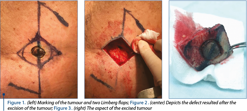
Considering the history of the lesion, the patient was admitted at the hospital, and after regular investigations (blood samples and EKG) he was planned for surgery. The surgery implied a circular incision with the electric cautery followed by another incision placed on the exterior of the previous one performed with a scalpel and the excision of the tumour with subcutaneous fat with 20 mm margin (Figures 1 and 2). The resulting defect was then covered by creating one local Limberg flap. After minutious haemostasis and adequate cleaning of the wound, the suture was performed in anatomical layers (Figure 4).
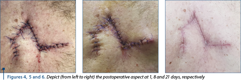
In the following days, the patient was assessed and dressings were performed daily. The evolution was favorable, with the wounds healing nicely, without any complications. On the third day, the patient was discharged from the hospital with the recommendations of continuing the antibiotherapy, the administration of pain killers (if needed), and follow-up at 7 and 14 days.
The histopathology result, which was ready 21 days after the operation, confirmed the initial suspicion and had the result of malignant melanoma with Clarck III and Breslow index >0.76 mm (approximately 1.3 mm) and complete excision of the tumor.
Discussion
Malignant melanoma is not a rare disease in Romania. Being a serious illness, very aggressive and with potential of bad prognosis, it is of the outmost importance that is diagnosed as fast as possible, thus increasing the chances of survival(1).
Several studies have been performed on this subject, and in one of these Osterlind found that some characteristics influence the evolution of the disease, with women and younger patients having better prognosis and survival rates than men, and older people and better localized tumours being less aggressive than the disseminated ones(1,2).
The patient from our study had a localized lesion in the instercapullary region, but had higher risk, being an older-aged male.
Also, malignant melanoma seems to be more frequent in white people, as well as in Africans and others(3,4).
An important aspect is the classification of the malignant melanoma. Over the years, several classifications for malignant melanomas have been proposed, which include the Breslow, Clark and TNM classifications (the last being the most elaborate).
The more summarized versions are the first two, with Breslow indicating the depth of the lesion and offering a prediction of the future evolution, while Clark being used to describe mostly the thin forms of melanoma(5,6).
Breslow classification:
- Thickness of 0.75 mm or less
- Thickness of 0.76-1.5 mm
- Thickness of 1.51-4 mm
- Thickness greater than 4 mm.
Clark classification:
Level I – involves only the epidermis (in situ melanoma).
Level II – invades papillary dermis, but not the papillary-reticular interface.
Level III – invades the papillary dermis up to the interface, but without the reticular dermis.
Level IV – invades the reticular dermis, but not the subcutaneous tissue.
Level V – invasion into the subcutaneous tissue.
Conclusions
Malignant melanoma is gaining a rapidly increasing incidence yearly worldwide. It remains an important and challenging issue because of the subclinical micrometastases that it may generate and which can affect the other organs in the same time in which the primary lesions is being treated. Hence the importance of clinical trials and screening in evading complications(1,6).
Conflict of interests: The author declares no conflict of interests.
Bibliografie
- Yagi KI, Wani HU, Tinguria MB, Al-Qahtani DM. Malignant melanoma of the skin: a case report from the eastern region (Dammam Central Hospital), Saudi Arabia and epidemiological review. Journal of Family & Community Medicine. 2000; 7(2):67-71.
- Osterlind A, Kjems E. Survival of Danish Cancer Patients 1943-1987 M.M.of the Skin. APMIS Suppli Danish Cancer Society Epidemiology. 1993; 3:149–55.
- Katsambas A, Nicolaidou E. Cutaneous Malignant Melanoma and Sun Exposure. Recent Developments in Epidemiology. Arch Dermatol. 1996; 132(4):444–50.
- Mason MD, Allman R, Quibell M. Adhesion molecules in melanoma – more than just superglue. JR Soc Med. 1996; 89(7):393–5.
- American Joint Committee on Cancer. Malignant melanoma of the skin. American Joint Committee on Cancer: AJCC Cancer Staging Manual. 5th ed.1997; Philadelphia, Pa: Lippincott-Raven, 163-70.
- Neligan CP. Plastic Surgery, 3rd edition. Elsevier Saunders.2013.
Articole din ediţiile anterioare
REVIEW | Ediţia 2 51 / 2020
Managementul tratamentului şi urmărirea pacienţilor cu melanom malign în perioada pandemiei de COVID-19
Daniela Zob, Cornelia Niţipir, Prof. Dr. Laura Mazilu, Gabriela Niculai, Alina Moldovan, Lidia Kajanto, Andra-Iulia Suceveanu, Radu Florin, Silviu VOINEA, Viorel Savin, Amalia Constantinescu, Alexandru Grigorescu, Raluca Ioana Mihăilă, Conf. dr. Dana Lucia Stănculeanu
Melanomul este o problemă de sănătate publică, prin incidenţa sa în creştere şi prin vârsta mică la diagnostic. Există trei subtipuri de melanom...
19 mai 2020
CASE PRESENTATION | Ediţia 1 66 / 2024
Malignant melanoma of the leg – challenges. A case report
Moraru Mihaela , Goga Iolanda , Ilie Maria-Andreea, Stănculeanu Dana-Lucia , Gheorghe Adelina-Silvana , Dumitrescu Elena-Adriana , Zob Daniela-Luminiţa
Melanomul malign cutanat este al 15-lea cancer ca frecvenţă în rândul femeilor, iar incidenţa şi mortalitatea asociate melanomului malign continuă ...
27 martie 2024
CASE PRESENTATION | Ediţia 1 66 / 2024
Diffuse large B-cell lymphoma and metachronous metastatic renal cancer – a case report
Goga Iolanda , Cioc Enache , Boghici Elena-Monica , Moraru Mihaela , Nichita Monica-Adriana, Dumitrescu Elena , Pirlea Andrei-Daniel , Zob Daniela-Luminiţa
Dintre tumorile tractului urinar, cancerul renal este cel mai frecvent, iar în ceea ce priveşte subtipul histopatologic, cel cu celule clare ocupă ...
27 martie 2024
REVIEW | Ediţia 4 57 / 2021
Melanomul malign ocular – corelaţia evoluţiei clinice cu parametrii biologici şi imunologici
Păsărică Mihai Adrian, Dragosloveanu Christiana Diana Maria, Maria Iuliana Gruia, Gabriela Murgoi, Schmitzer Speranţa, Alexandru Grigorescu
Melanomul malign, deşi relativ mai puţin frecvent comparativ cu restul neoplaziilor cutanate, este responsabil de cele mai multe fatalităţi prin c...
20 decembrie 2021