Rapid maxillary expansion is defined as the release of medio-palatine suture using an orthopedic forces. The role of this procedure is to expand the upper jaw in order to achieve the broadening of the upper arch. This study was initiated to quantify the effects of disjunction and post- treatment bone changes, after an adequate contention which lasted for three weeks. This study is carried on laboratory animals (common breed rabbit) to determine tensile strength and the elasticity modulus of biological materials used in orthdodontics. The results of the study are consistent with those reported in the literature reference.
INTERDISCIPLINARE
Calculul indicilor utilizaţi pentru cuantificarea expansiunii rapide de maxilar
Calculations used for assessment of rapid maxillary expansion
Ruxandra Bartok,
Bogdan Dimitriu,
Constantin Vârlan,
Radu Stanciu,
Georgiana Moldoveanu,
Paul Gagniuc,
Ileana Suciu,
Irina-Maria Gheorghiu,
Loredana Mitran,
Mihai Mitran,
Ioana Suciu
First published: 10 martie 2016
Editorial Group: MEDICHUB MEDIA
DOI: 10.26416/Orl.30.1.2016.525
Abstract
Rezumat
Expansiunea rapidă de maxilar este definită ca eliberarea suturii medio-palatine folosind forțe ortopedice. Rolul acestei proceduri este de a dilata maxilarul superior pentru a obține lărgirea arcadei maxilare. Studiul de față este realizat pentru a cuantifica efectele disjuncției și modificarile de la nivelul osos maxilar, ulterioare utilizării acestei metode de tratament ortodontic. Cercetarea este realizată pe animale de laborator (iepure de rasă comună) pentru a determina rezistența la tracțiune și modulul de elasticitate a materialelor biologice utilizate în ortodonție. Rezultatele studiului sunt în concordanță cu cele raportate în literatura de referință.
Calculul indicilor utilizaţi pentru cuantificarea expansiunii rapide de maxilar
Rapid maxillary expansion (RME) has been proposed by Angle since the 21st century, in order to correct jaw constriction(2,4). RME is a routine procedure in orthodontics, with the primary goal of expanding the narrow maxillary arch. Large forces used during the activation of an expansion screw split the intermaxillary suture(10,11,13), thus increasing in width both the basal bone and the width of the dental arch perimeter(1). Since 1920, Mesnard demonstrated radiologically that medio-palatine suture may be opened by using the expansion device and the resulting space would be filled with bone during 4-6 weeks(5,6,12).As far as the tensile testing of materials regards, of biological materials in this case, this is an adequate method to determine their behavior on axial tensile loads. The data from the tests are used to determine tensile strength and the elasticity modulus.
The tensile stress is determined by an applied load, which extends the material on the forces’ axis application.
Stress voltage is a condition caused by pregnancy, which extends applicas forces.
Uniaxial stress expresses through:
where F is the force acting on area A (m2). The approach to linear elastic fracture mechanics is to estimate the amount of energy required to raise a pre-existing crack in a brittle material.
Where E is the Young’s modulus (the elasticity modulus) - specific for the tested material, γ is the surface energy per unit area of the crack and a is the crack length on side, whereas 2a represents the crack length inside a flat surface.
The expression
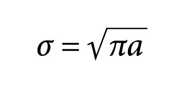
The uniform uniaxial stress
A parameter called the stress intensity factor (K) is used to determine the tensile strength of most materials. Stress intensity factor (K) for a 2a crack length(14,16) at right angles in an infinite plane, subject to a uniform force field stress (σ) is:The medio-palatine suture could be considered as a pre-existing crack.Therefore, to calculate the necessary forces(7) in RME we consider a crack centrally located in a finite plate.
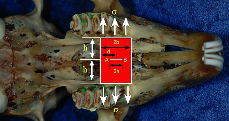
Initiated crack on a middleflat surface under uniaxial stress conditions
If the crack is centrally located along the width (d≠b), the stress intensity factor at the location A has the form (Figure 1):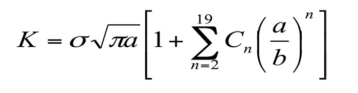
where C n factors can be found in tables for various values of d. A similar expression can be found for the crack tip B (Figure 1).
Material and method
Assuming the occurence of medio-palatine disjunction to be initiated in the middle of a flat surface, under uniaxial stress conditions (as detailed in Figure 1) the expansionat of the medio-palatine suture was measured, for a range of experimental animals, which were prepared according to a predetermined protocol.For the current study were used a total of 12 rabbit dried skulls.
They emerged from an in vivo study on 8 common breed rabbits, following the rapid disjunction; the disjunctor remained anchored to the side zone for a period of 24 days.
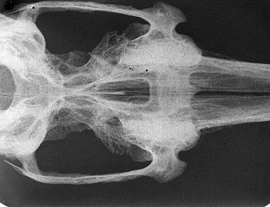
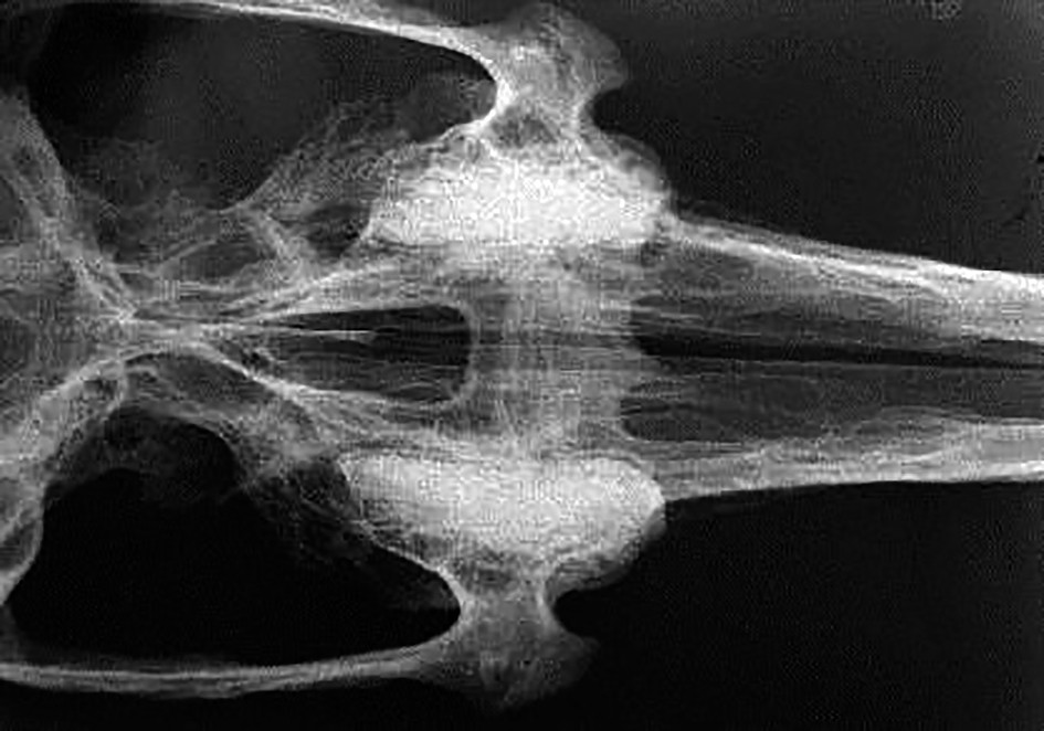
After euthanasia, according to the working protocol, followed the procedure of posting organic substrate from the support of animal bone, using a concentrated solution of NaOCl 9% at a temperature of 55⁰C. After drying, it resorted to measurements so that was taken into account intermolar distance on first maxillary molars (Figure 2, Figure 4).
Four dry skulls served as control to benchmark expansion coefficient obtained at a distance of 24 days, after rapid disjunction, enabling the disjunctor applied over this period to maintain the result and conservation reossification trend (Figure 3).
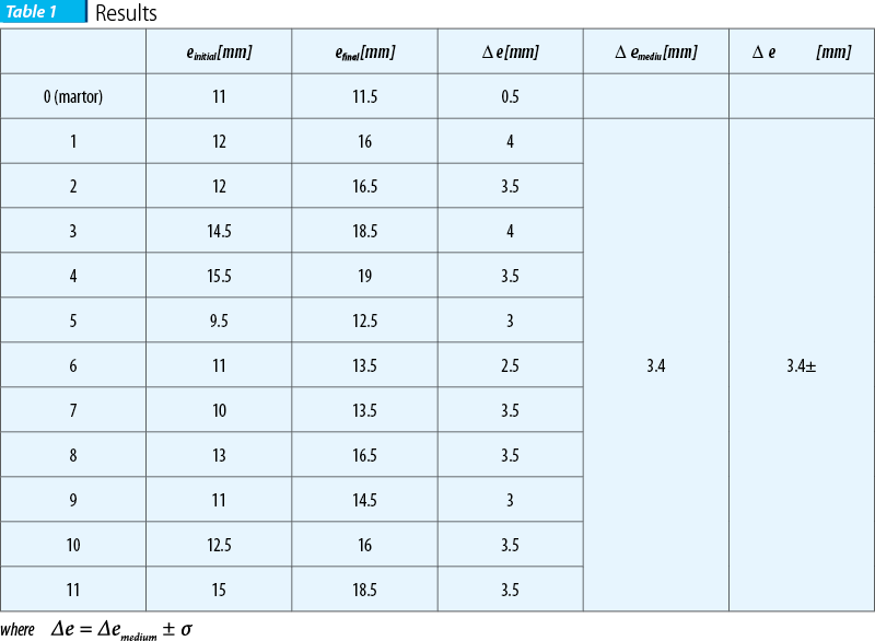
We further note with e (where e = 2h) the medio-palatine expansion. The results obtained are shown in Table 1.

Discussion
The study was initiated to quantify the effects of disjunction and post-treatment bone changes, after an adequate contention which lasted for three weeks. For the statistical data processing it was assumed that measurements are described by a Gaussian normal distribution. All measurements were carried out by two operators, and subsequent errors were calculated by comparing the first with the second sample to give an average of bone growth of 3.4 mm transversely to the segment being analyzed. All measurements of random error coefficients are placed in acceptable limits. In all investigated cases, the opening of the medio-palatine suture occurred at an important ratio in the anterior segment rather than in the posterior segment of the suture(8,9,15).
Conclusions
After analyzing the results, recorded on dried skull rabbit group, post-disjunction and contention, it was found that all coefficients resulting from linear intermolar transversal measurements tended to be significantly increasing.It is interesting to note that these results are consistent with those reported in the literature reference.
Bibliografie
1. Adkins MD, Nanda RS, Currier GF: Arch perimeter changes on rapid palatal expansion. Am J OrthodDentofacialOrthop. 1990;97(3):194–199.
2. Angle EC.: Treatment of irregularities of the permanent or adult teeth. Dent Cosmos 1860;1:540-4.
3. Bo Hou, Naomi Fukai, Bjorn R. Olsen.: Mechanical force-induced midpalatal suture remodeling in mice Bone 40 (2007) 1483 – 1493.
4. Byloff FK, Mossaz CF.: Skeletal and dental changes following surgically assisted rapid palatal expansion. European Journal of Orthodontics 2004:26:403-409.
5. Cameron CG. Franchi L, Baccetti T, McNamara JA.: Long-term effects of rapide maxillary expansion: a posteroanteriorcephalometric evaluation. Am J Orthod Dentofacial Orthop 2002;121:129-35.
6. Charezinski M., Balon-Perin A., Deroux E., De Maertelaer V., Glineur R.: Transverse maxillary stability assisted by a transpalatal device: A retrospective pilot study of 9 cases, Int. J. Oral Maxillofac. Surg. 2009; 38:937-341.
7. Cozza P, Giancotti A, Petrosino A.: Rapid palatal expansion in mixed dentition using modified expander: a cephalometric investigation. J Orthod 2001;28:129-34.
8. Davidovitch M, Efstathiou S, Sarne O, Vardimon AD.: Skeletal and dental response to rapid maxillary expansion with 2- versus 4-band appliances. American Journal of Orthodontics and Dentofacial Orthopedics 2005;127: 483-92.
9. Djasim UM, Hekking-Weijma JM, Wolvius EB, van Neck JW, van der Wal KG.: Rabbits as a model for research into craniofacial distraction osteogenesis. British Journal Oral and Maxillofacial Surgery 2008;46:620-4.
10. Haas AJ.: Rapid expansion of the maxillary dental arch and nasal cavity by opening the midpalatal suture. Angle Orthod 1961;31:73-90.
11. Hayes J.L.: Rapid maxillary expansion, Am J Orthod Dentofacial Othop 2006;130:432-3.
12. Mesnard L. Immediate separation of the maxillae as a treatment for nasal impermeability. Dent Rec 1929;49:371-372.
13. Oliveira NL. Da Silveira AC, Kusnoto B, Viana G.: Three-dimensional assessment of morphologic changes of the maxilla: a comparison of 2 kinds of palatal expanders. Am J Orthod Dentofacial Orthop2004;123:354-62.
14. Parr JA, Garetto LP, Wohlford ME, Arbuckle GR, Roberts WE, Implant-borne suture expansion in rabbits: a histomorphometric study of the supporting bone. J Biomed Mater Res 1999;45:1-10.
15. Pearce AI, Richards RG, Milz S. Schneider E, Pearce SG. Animal models for implant biomaterial research in bone. a review. Eur Cell Mater 2007;13:1-10.
16. Podesser B, Williams S, Crisman AG, Bantleon HD. Evaluation of the effects of rapid maxillary expansion in growing children using computer tomography scanning: a pilot study. Eur J Orthod 2007;29:37-44.