Background. Extraskeletal Ewing’s sarcoma is a rare tumor of mesenchymal origin, which is histologically similar to primary osseous Ewing’s sarcoma. It has been reported a low incidence in the head and neck.
Methods. We report the case of a patient with extraskeletal Ewing’s sarcoma presenting with a lateral and posterior left sided neck mass. Magnetic resonance imaging was useful in preoperative evaluation. The treatment consisted in radical resection, and the histological analysis completed the diagnosis.
Results. The postoperative management consisted in a combination of chemotherapy for systemic control and irradiation for local control. PET-CT was useful to evaluate the treatment’s efficacy.
Conclusions. Extraskeletal Ewing’s sarcoma is a rare and aggressive tumor. A very few cases of cervical tumors were reported. Radical resection followed by radio-chemotherapy has good results in the treatment of this rare tumor.
Sarcom Ewing extrascheletal sub forma unei mase tumorale la nivelul gâtului - prezentare de caz
Extraskeletal Ewing’s sarcoma presenting as a neck mass - case report
First published: 07 iunie 2017
Editorial Group: MEDICHUB MEDIA
DOI: 10.26416/Orl.35.2.2017.795
Abstract
Rezumat
Introducere. Sarcomul Ewing extrascheletal este o tumoră rară, de origine mezenchimală, similară din punct de vedere histologic cu osteosarcomul Ewing. Este o tumoră rar întâlnită la nivelul capului și gâtului.
Metode. Autorii prezintă un caz de sarcom Ewing extrascheletal sub forma unei tumori laterocervicale stângi situate posterior de mușchiul sternocleidomastoidian. Examenul RMN a fost util în evaluarea preoperatorie. Tratamentul a constat în excizia radicală a tumorii laterocervicale, urmată de examen histopatologic.
Rezultate. Postoperator s-au efectuat chimioterapie pentru controlul sistemic și radioterapie pentru controlul local. Evaluarea eficienței schemei terapeutice a fost realizată prin examen PET-CT.
Concluzii. Sarcomul Ewing extrascheletal este o tumoră rară, cu evoluție agresivă. În literatură au fost raportate puține cazuri de sarcoame Ewing extrascheletale prezentate sub forma unor tumori laterocervicale. Excizia radicală urmată de radiochimioterapie are cele mai bune rezultate în tratamentul acestor tumori.
Introduction
Extraskeletal Ewing’s sarcoma (EES) is an uncommon tumor occurring mostly in children and young adults. It was first recognized by Tefft et al. in 1969, who reported four cases of paravertebral soft tissue tumors that histologically resembled with Ewing’s sarcoma(1).
The most common sites for EES are trunk, extremities, retroperitoneum and head and neck in about 18% of cases(2,3,4). According to Chao et al., there are only 5 out of 118 cases of EES located in head and neck region(5). In head and neck, EES is usually identified in nasal or oral cavities, sinuses or soft tissues(6). EES is characterized by an aggressive course and high rate of recurrences. The treatment consists in a combination of surgery, radiotherapy and chemotherapy.
Case report
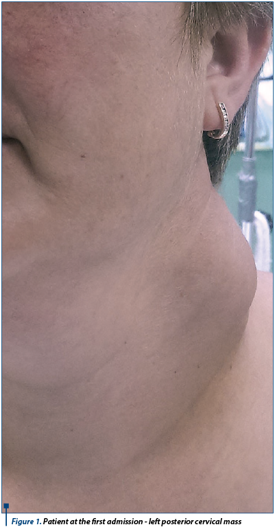
A 39-year-old female was admitted in ENT Department with a 2-month history of painless posterior left laterocervical mass with rapid evolution (Figure 1).
The patient’s medical history showed systemic lupus erythematosus with chronic administration of 10 mg of prednisolone per day and cervical cancer 6 years ago, treated by radical surgery followed by radiotherapy.
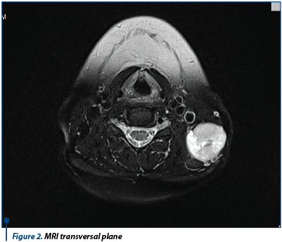
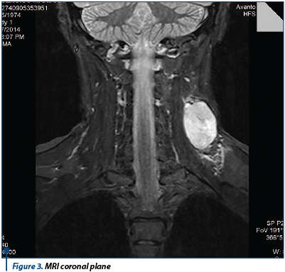
The videofibroscopy of rhinopharynx, hypopharynx and larynx was normal. The MRI showed a posterior laterocervical solid mass, with a maximum diameter of 8 cm (Figure 2 and Figure 3).
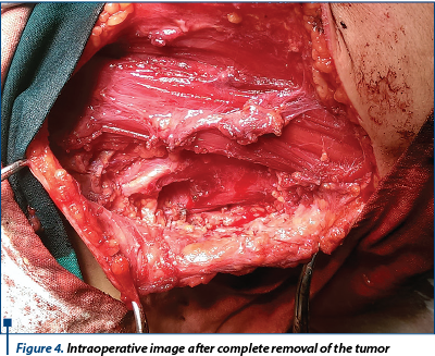
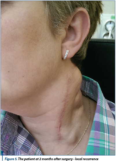
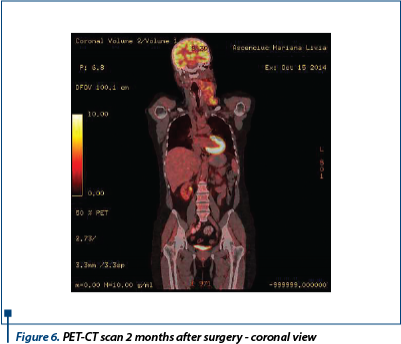
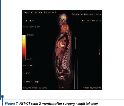
The patient underwent surgical excision followed by histopathological exam (Figure 4). The first histopatological exam showed non-Hodgkin lymphoma, but immunohistochemical studies revealed that tumor cells were positive for CD99 and FLI1 - highly suggestive for Ewing’s sarcoma. For unexpected reasons, the patient didn’t start postoperative radiotherapy, and at two months after surgery she presented in our department with local recurrence (Figure 5). This time, PET-CT showed very high metabolic activity in the neck mass, with no distant metastases (Figure 6 and Figure 7). The patient underwent 6 chemotherapy cycles with vincristine, doxorubicin and cyclophosphamide, followed by external radiotherapy.
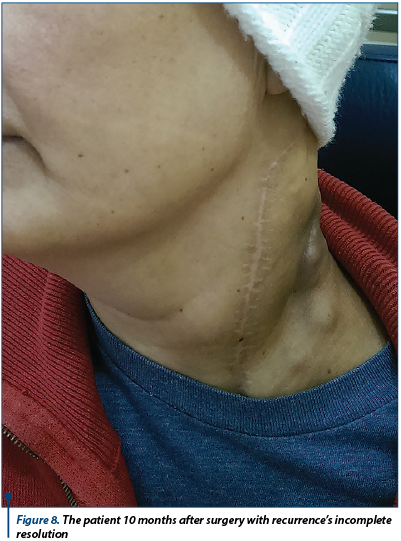
At 10 months after initial diagnosis, the patient presented with incomplete resolution of the tumor (Figure 8), severe radiomucositis (grade 4 WHO) and was admitted in the oncology department.
Discussion
The diagnosis of Ewing’s sarcoma was made by immunohistochemistry tests, showing the presence of CD99 and FLI1 on cell membrane.
EES in head and neck region must be differentiated from lymphoma, rhabdomyosarcoma, poorly differentiated salivary gland tumors, olfactory neuroblastoma, undifferentiated nasopharyngeal carcinoma and melanoma.
Case particularities
- The localization of EES in the neck is very rare.
- The patient’s age: most of the cases of EES were reported in patients no older than 30 years.
- Comorbidities - immunosuppression caused by chronic use of prednisolone could be an etiologic factor.
- Delay of the radiotherapy after surgery (2 months) caused tumor recurrence, EES being a very aggressive type of tumor.
Bibliografie
2. Hafezi S, Seethala RR, Stelow EB, et al. Ewing’s family of tumors of the sinonasal tract and maxillary bone. Head Neck Pathol. 2011; 5:8–16. doi: 10.1007/s12105-010-0227-x.
3. Shimada H, Newton WA Jr, Soule EH, et al. Pathologic features of extraosseous Ewing’s sarcoma: a report from the Intergroup Rhabdomyosarcoma Study. Hum Pathol. 1988; 19:442–453. doi: 10.1016/S0046-8177(88)80495-7.
4. Stuart-Harris R, Wills EJ, Phillips J, et al. Extraskeletal Ewing’s sarcoma: a clinical, morphological and ultra-structural analysis of five cases with a review of the literature. Eur J Cancer Clin Oncol. 1986; 22:393–400.
5. Chao TK, Chang YL, Sheen TS. Extraskeletal Ewing’s sarcoma of the scalp. J Laryngol Otol.2000; 114:73–75.
6. Windfuhr JP. Primitive neuroectodermal tumor of the head and neck: incidence, diagnosis, and management. Ann Otol Rhinol Laryngol. 2004; 113:533–543.